CBCT (Cone Beam CT) in Dentistry
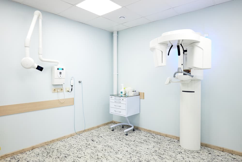
Cone beam computed tomography (CBCT) has brought a revolution in the world of imaging in dentistry. It is the most advanced imaging technique.
Unlike traditional X-rays, which offer only limited two-dimensional views, cone beam computed tomography (CBCT) produces a comprehensive three-dimensional view, capturing fine details with exceptional accuracy. It offers highly precise and detailed anatomical information.
This technology allows dentists to assess the width, height, volume, and density of bone, improving diagnosis, which ultimately improves patient care.
It accurately defines the width, height, density, and volume of bone.
Embracing CBCT not only elevates the standard of dental imaging but also ensures that patients receive the most informed and precise treatments available.
Introduction to CBCT
Cone beam computed tomography (CBCT) has transformed dental imaging. It has changed how dentists visualize various structures within the oral cavity. It provides a bigger picture of all dimensions of anatomical structures, including;
- Cross-sectional view.
- Axial view.
- Panoramic view.
The most common use of CBCT is when placing a “dental implant” in patients.
You can use it for other uses as well. It can be used in “endodontics”; in root fracture, especially in “vertical root fracture” that cannot be identified on an X-ray, or in patients having very “complex anatomy”, whether the patient has calcified canals, and if the number of canals cannot be identified otherwise on a conventional X-ray.
In oral and maxillofacial surgery, it is used to check the extent of fracture and the damage.
It can also be used in orthodontics to check for the placement of canines.
Benefits of CBCT:
1. Bone Graft/ Sinus Lift:
Can be used for the assessment of the need for a bone graft or sinus lift.
2. Vital Anatomical Structures:
Can be used to measure the distance between vital anatomical structures.
3. Implant Placement:
Plan optimal implant placement and angulation.
4. Minimizing surgical time:
Minimize surgical time
5. Bone's Width and Height:
Used for the evaluation of the alveolar bone's width and contour.
6. Selection of Implant:
Selection of the most suitable implant model and size.
7. Accuracy:
CBCT reproduces highly precise dental imaging. It is unmatched when compared to any other imaging technique in dental practice. It is a benchmark imaging technique, a benchmark imaging technique especially when considering performing any surgical procedure in dentistry.
CBCT allows for precise dental implant placement by defining the bone density and volume, which are critical for implant success.
8. Lower radiation dose:
CBCT gets the edge as it minimizes the dosage of radiation, making it a more preferable option when evaluating anatomical structures of the oral cavity.
It also helps in tracking bone regeneration in implant sites.
9. Fast
CBCT scan gives you a super speedy turnaround, saving you time as a patient and a dental practitioner.
It is a quick and easy way to evaluate and provides detailed information within just a few seconds time span, hence speeding up the decision-making procedure as well as treatment planning.
10. Versatile
The scope of CBCT scans extends across a wide range of dental issues. They are instrumental in diagnosing and managing jaw tumours by providing a clear view of the extent and location of the growth. For orthodontic assessments, CBCT scans can show the position of unerupted or misaligned teeth, aiding in more accurate treatment planning.
CBCT (Cone beam computed tomography) is an incredible diagnostic tool that aids in diagnosing various complex dental issues with exact precision.
It helps in all fields of dentistry, whether it be endodontics, orthodontics, oral and maxillofacial surgery (OMFS), or prosthodontics.
They provide a clearer view of jaw tumors. Defining the extent and location of the tumor for efficient removal by the surgeon.
Differences between CBCT and traditional CT scans:
Radiation Dose Comparison
CBCT scans have an important advantage over conventional CT scans by minimizing the dose of radiation.
Image Quality
Both CBCT scans and CT scans provide 3D imaging; CBCT produces excellent image quality.
Economical:
CBCT is pocket-friendly, while CT scans are more expensive comparatively.
Radiation dose comparison:
CBCT Image Interpretation
Understanding CBCT Images
Interpretation of CBCT images requires detailed knowledge of the anatomical structures of the oral cavity. With the help of CBCT scans, dentists can assess various factors, including bone volume and density, alignment of teeth, and nerve location, to deliver appropriate treatment to the patient. It helps in diagnosing the condition earlier to provide effective treatment.
Common Findings in CBCT Scans
CBCT scans can reveal conditions such as;
- Hidden root fractures.
- Impacted teeth.
- Oral Cysts.
- Bone density.

